
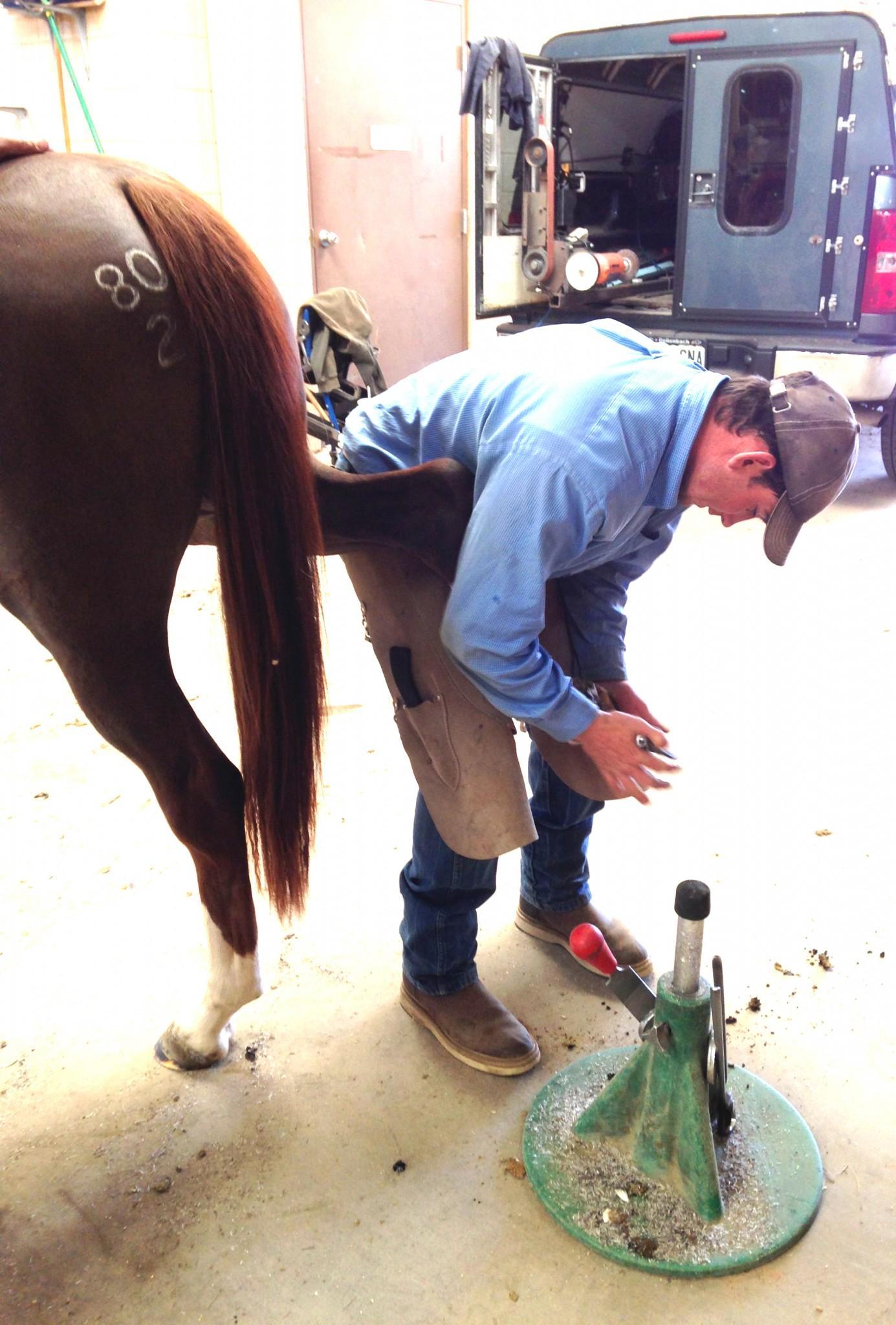
There’s an old saying in the equine industry, “no foot, no horse.” But I found out this week in farrier practicum it should really be “no middle finger, no horse.” Yes, that’s right. Horses walk on their middle fingers in their forelimbs and their middle toe in the hind limbs. Sometimes people refer to them as toe-walkers. That’s really weird to think about.
Can you imagine putting 60% of your body weight on your middle fingers? Not even in yoga class. Try it on the nail of your middle fingers? Ouch! I don’t know about you, but I don’t think I could stand it.
The hoof of a horse is an incredible structure. My toe-walking friends accomplish amazing feats of athleticism and power atop such small objects compared to their weight and body size. But proper care of the hoof is key to the horse’s health a ability to do all we ask them to do as athletes. Mess with the hoof and you’ve got problems.
The hoof capsule, the part we compare to our nails, is made up of epithelial skin cells that have been keratinized. There are a lot of layers in the hoof wall. The part we can see from the outside is called the hoof varnish. Like a fingernail that grows slowly from the cuticle, the hoof grows from the coronary band down to the ground.
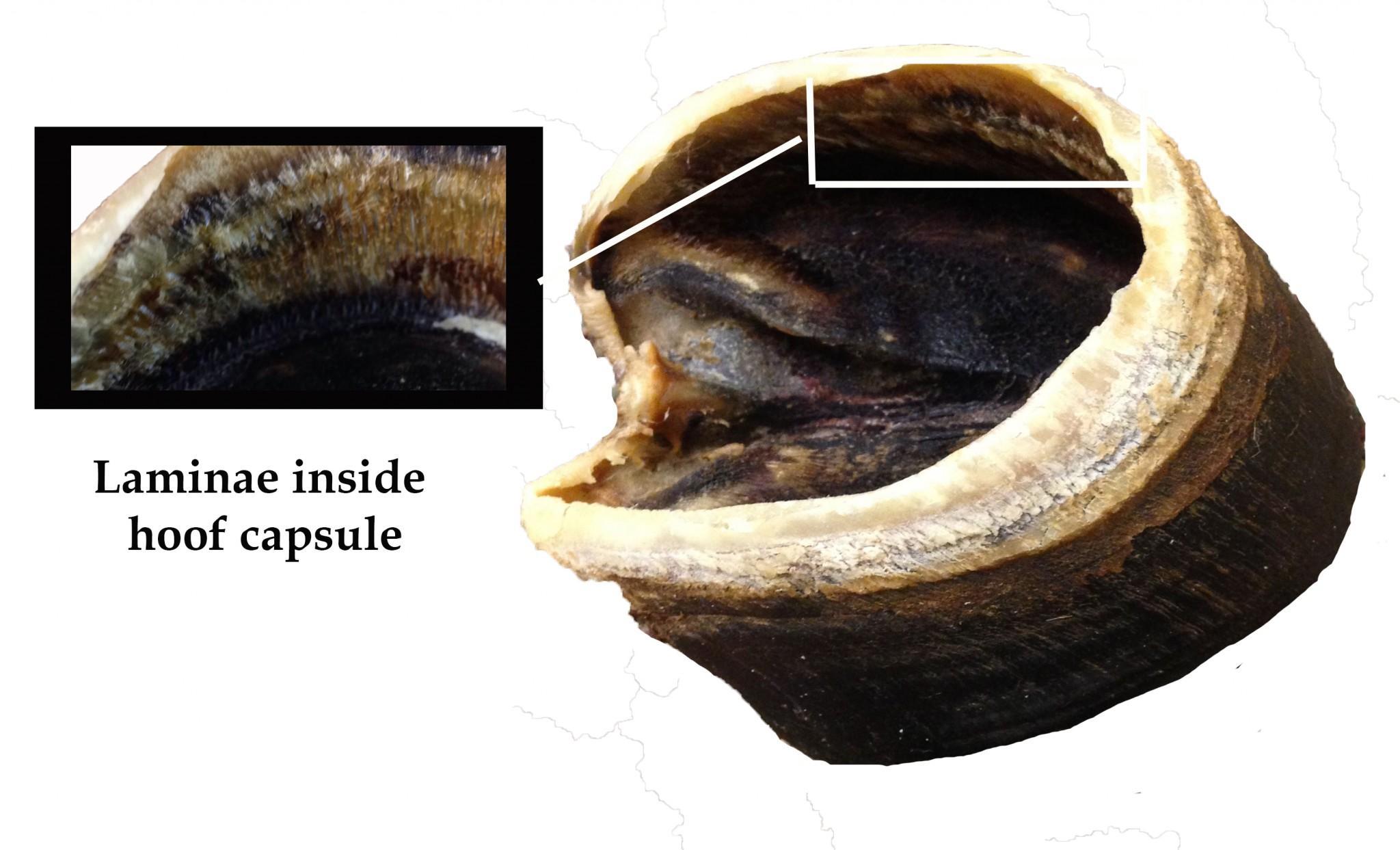
Inside the hoof wall, past the hoof varnish there is a section of interlocking fingerlike projections called the horny laminae and sensitive laminae. This is the point of attachment between the hoof wall and the tip of the middle finger in the forelimb, which in the horse is called the coffin bone, P3 or third phalanx.
Don’t worry I’m not going to go super anatomist on you. I just kind of can’t believe that the tissue holding the hoof onto the bone is as strong as it is. If a horse is carrying about 60% of its body weight on the front limbs alone, let’s say in an 1100-pound horse, that’s about 660 pounds. Gosh! I can’t imagine a human fingernail having that kind of strength to stay attached.
In dissection when you see the hoof capsule separated from the boney structure, the horny laminae kind of look like whale baleen with lots of ridges. It’s quite amazing to ponder how this system works.
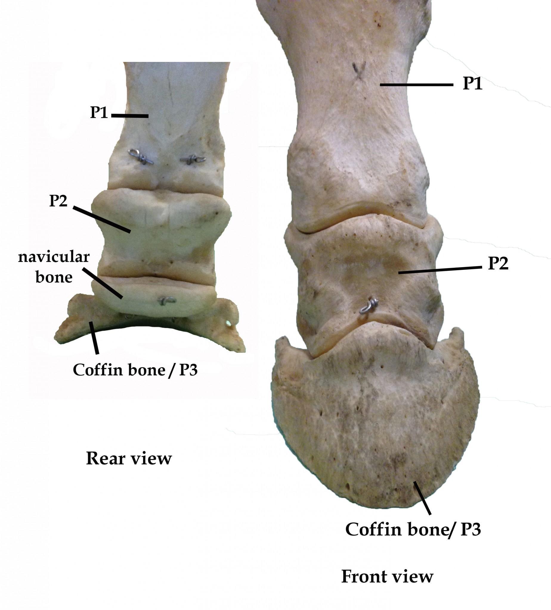
The hoof capsule, besides housing the coffin bone, also accommodates the distal sesamoid bone, which is also called the navicular bone. Look, I don’t know why all these bones have multiple names. If I find out I’ll let you know, but for now let’s talk about the function of this little bone. It acts like a pulley with the deep digital flexor tendon sliding across it attached to the underside of the coffin bone. The capsule also contains the digital cushion.
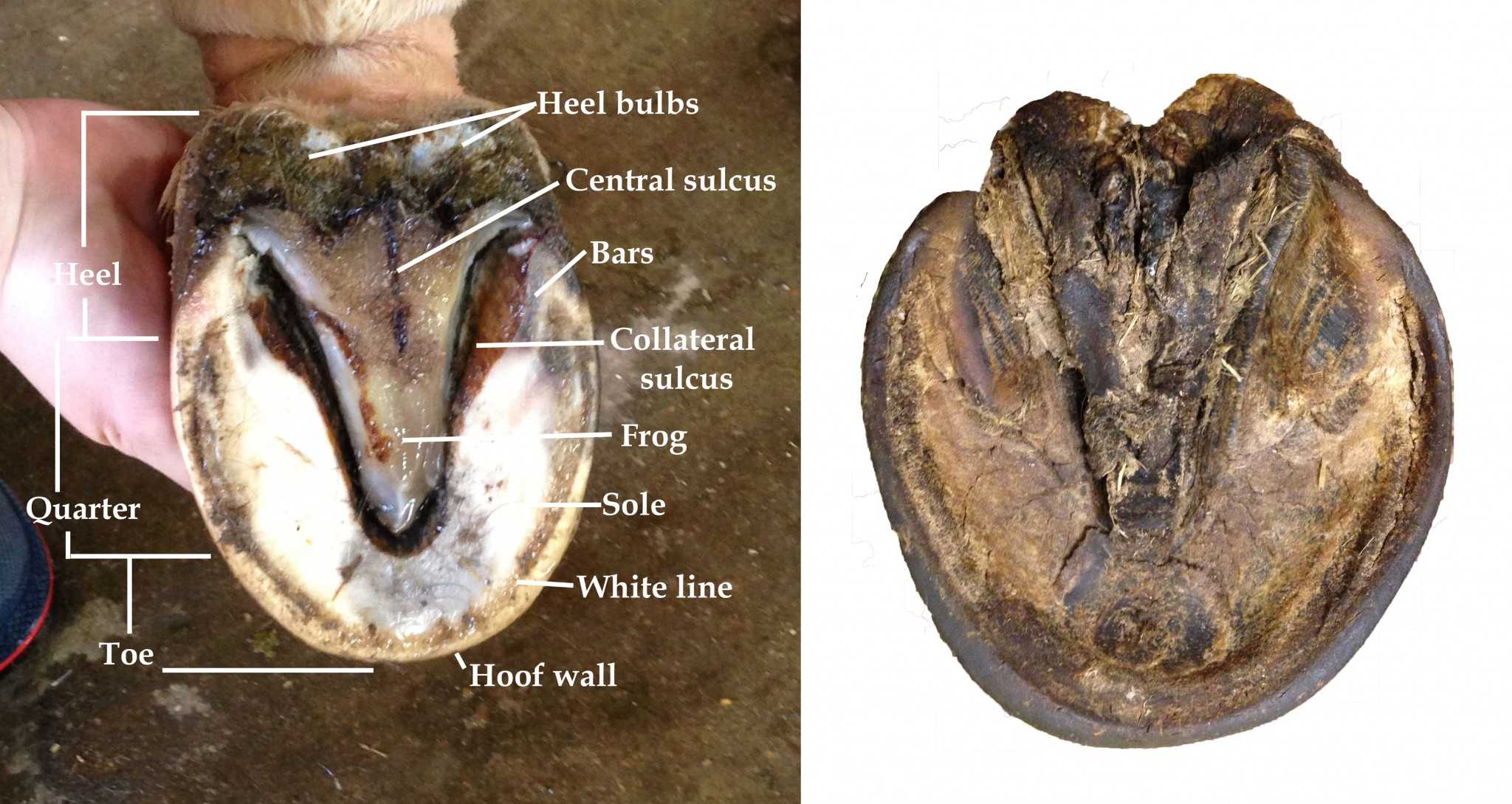
When you look at the hoof from below, you see the wall, an area called the white line, the sole, a triangle-shaped area called the frog, the bars, collateral sulcus along the edge of the frog, the central sulcus of the frog, and the heel bulbs. But the really cool thing about looking at the hoof either from the bottom or from the side is understanding where the coffin bone is inside.
When you divide the bottom of the hoof into four equal parts, the intersection is the point of balance for the horse, known as Duckett’s dot. It should be in front of the point of the frog. This means that the coffin bone is partially bisected by the imaginary line, but pretty far forward from what you might have thought it would be inside the hoof capsule.
Another really amazing thing is that the hoof is not a hard immobile structure. It actually expands when the horse’s weight is on it. The expansion and contraction of the hoof, including the frog helps move blood around in the bone and tissue of the hoof capsule.
We need the farrier to perform regular trims to help keep the hoof in proper balance. To make sure the angle of the hoof is set to an angle matching the pastern joint. This makes it so the coffin bone is sitting properly inside the hoof capsule, and there is no undue strain or rotation on the joints because of improper alignment. He also makes sure the sole and frog can perform their important roles. The farrier may be the first person to diagnose lameness. Through his experience he can guide you through the next steps including when to seek veterinary attention.
When this whole elegant system is in alignment, the horse can move in true freedom. So you can imagine anything that interferes with this system can really cripple a horse. Hooves can get infected, have abscesses, diseases, or laminitis which is inflammation of those little interwoven fingers holding the hoof wall to the bone. In severe cases laminitis becomes founder. This is where the laminae are actually detaching and the tension of the deep digital flexor tendon is pulling the coffin bone away from the hoof wall.
I can’t really explain why I get so excited about studying equine anatomy. Human anatomy was a chore for me. Maybe it’s because the horse is so majestic when it moves. Somehow it gives me even greater joy when I watch a horse at work or play to know how complicated it is to be a toe-walker while looking so simple on the outside.